


iSurf BrainView app for iPhone and iPad
iSurf BrainView is a Brain MRI tutor based on the MRI automatic segmentation produced by FreeSurfer.
FreeSurfer is a software package developed by investigators at the Athinoula A. Martinos Center for Biomedical Imaging.
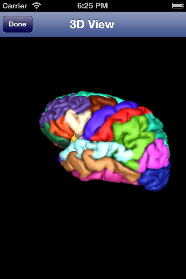
This App uses its automatic segmentation to produce an automatic atlas of neuroimaging based on T1 MRI Images.
A great tool for teaching brain MRI and for learning neuroanatomy.
Pros and cons of iSurf BrainView app for iPhone and iPad
iSurf BrainView app good for
The application is very useful to those starting in neuroscience.
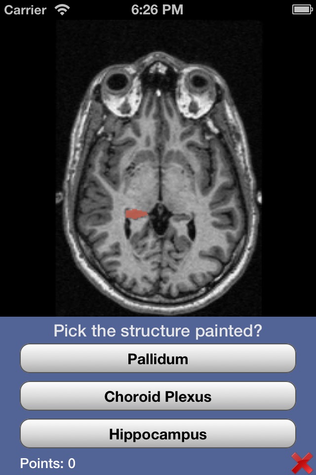
I enjoyed the snatomy quizzes and the apo really improved my orientation in the basal ganglia.
This app is a great way to study gross neuroanatomy by looking at and reading about structures generated by MRI. It is a good companion to apps such as Brain Tutor HD and 3D Brain.
I kind of like this app. It shows you a lot of things about the brain. Its may be one of the best medical apps ever!
A perfect app that allows me to touch up on some neuroanatomical regions when in the lab. Amazing tool for any researcher or analyst.
This helps beginners learn basic brain structure. Very good application. If possible, more info about sub cortical divisions are expected, such as hypothalamus, hypophysis, etc. etc.
Some bad moments
I do neuroimaging analyses for a living, so Im delighted to see tools being developed for the ipad/ipod. Thankyou!
That said, I have some real concerns:
1) I crashed the app 6-8 times in about 5 minutes on the ipad. It seems less finicky on the ipod (4th gen)...where I have yet to crash it.
2) The double arrow buttons do the same thing as the single arrow buttons on the ipad and ipod. That cannot be your intention.
3) The documentation appears to be for the full version of freesurfer. I have found no documentation for the app itself. Most folks would appreciate a little documentation
==================
Missing Documentation:
You can scroll through MRI slices in any of 3 orientations: sagittal, axial or coronal.
By selecting the little overlapping colored boxes at the bottom you can segment the brain (i.e., identify/colorize different tissue types and structures).
By touching the brain image in various locations you will see coordinates (lower right) and the name of recognized structures (upper left).
==================
Requests:
1) You include only 2 coordinates and no quantitative indication of which slice we are on...why not all 3 coordinates?
2) On the iphone/ipod touch, itd be nice to zoom.
3) Id like to know what this image is...it looks like an individual....is it in standard space? Is that talairach or MNI (i.e., the coordinates are more valuable if they are standard coordinates...but there is no indication of what they are).
4) Is there a possibility that we can upload our own structural images? Is that something you have planned for the future? Is this handling a real neuroimaging format like dicom or nifti...or is this a bunch of tiffs or what...very curious.
I hope you will continue to develop this.
High quality axial, coronal, and sagittal MR images of the brain. The slider bar lets you move smoothly from slice to slice; the animation button does this automatically and is helpful for visualizing how structures like the caudate migrate through the brain. This helps to get a 3-D sense. The ability to fill distinct structures with color is helpful but many important structures are poorly labelled. For example, on axial the optic nerve is an "unknown structure"; the internal capsule is "cerebral white matter". It would also be a nice addition to have a list of structures to choose from that would then be indicated on the scans.
I dont regret buying it though and will keep it on my iPad.
Nice scrolling in the coronal and sagittal planes to get an idea as to white matter tract courses. Not enough fine detail regarding neuroanatomy. For instance, instead of the generic "brainstem" there should at least be medulla, pons, midbrain... And some of the obvious things like vermis of the cerebellum, red nucleus, internal capsule, etc should be labelled. This app needs some more work.
My bad...you tap on any structure to see a label for the anatomy. Great app!!!
I LOVE this app!! This is exactly what i need for my research! Thank you so much for your work. If you could include brodman area, that would be great! Also another suggestion is to keep the point fixed on three different views, it will help a lot as well. Is it possible to use a MNI atlas instead of a single subject?
Great for learning how some neural structures typically look on MRI.